
Published May 13, 2022

Victoria L. Mango
Department of Radiology, Memorial Sloan Kettering Cancer Center

Lars J. Grimm
Department of Radiology, Division of Breast Imaging, Duke University School of Medicine

Jennifer A. Harvey
Department of Imaging Services, University of Rochester Medical Center

Donna M. Plecha
Department of Radiology, University Hospitals Cleveland Medical Center

Emily F. Conant
Department of Radiology, Division of Breast Imaging, Perelman School of Medicine, University of Pennsylvania
Abbreviated breast MRI (AB-MRI) has been shown to maintain the high sensitivity of longer or full breast MRI protocols while decreasing operational costs. The clinical implementation of an AB-MRI program requires collaboration of multiple stakeholders, including administrative, operational, financial, technical, and clinical providers. Institutions must define patient eligibility and imaging protocols and monitor performance metrics to ensure high-quality patient care. The improved efficiency and maintenance of accuracy of AB-MRI may allow more women to access this important supplemental screening modality.
MRI is the most sensitive imaging modality for breast cancer detection [1], and its role as an adjunct to mammographic screening for women at high risk for breast cancer (i.e., a lifetime risk > 20%) is well accepted [2–5]. Although demographic and genetically based risk assessment models are often used to determine breast cancer risk, increased mammographic density increases breast cancer risk and decreases mammographic sensitivity through the obscuration of cancer by adjacent dense breast tissue [6]. Increasing awareness of the impact of increased breast density on patient outcomes has driven the adoption of state and federal legislation mandating that women be notified of their breast density. In response to this increasing awareness, there is growing interest in supplemental screening for breast cancer with breast MRI, which shows improved breast cancer detection for women with dense breasts and otherwise average risk [7]. To accommodate the increasing demand for supplemental breast MRI screening, abbreviated (or fast) breast MRI protocols have been developed that reduce costs, improve workflow, and increase patient access. This article provides details regarding the current literature supporting the utilization of AB-MRI for supplemental screening and the numerous administrative, operational, financial, technical, and clinical elements that must be considered in the implementation of a clinical AB-MRI program.
Review of Abbreviated Breast MRI Outcome Data
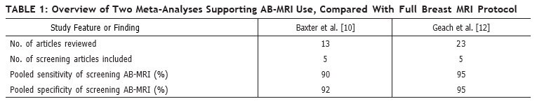
The first AB-MRI study, which was published in 2014 by Kuhl et al. [8], included 443 women with a “mildly to moderately increased risk of breast cancer.” The study showed that a 3-minute AB-MRI acquisition that consists of only one unenhanced and one contrast-enhanced sequence has diagnostic accuracy in breast cancer screening that is equivalent to that of a full breast MRI protocol. Multiple studies have since been published, including a prospective multicenter trial [9] and at least three systemic reviews or meta-analyses supporting the use of AB-MRI [10–13] (Table 1).
These studies confirm that the combination of mammography and MRI screening yields the highest cancer detection rate. In addition, cancers detected with MRI are more likely to be invasive and higher grade, compared with cancers detected with mammography [14], and therefore have the potential for greater impact on patient morbidity and mortality.
Because MRI offers improved cancer detection rates and can detect more clinically relevant disease, AB-MRI has been integrated into clinical practice at numerous institutions. Clinical implementation of an AB-MRI program requires consideration of patient eligibility requirements, imaging protocols, and appropriate performance metrics to ensure high-quality patient care.
Patient Selection and Screening Interval
At present, most supplemental screening with AB-MRI is reserved for women with dense breasts, given the superior sensitivity of breast MRI for cancer detection [15]. This recommendation is supported by multiple studies of women with dense breasts, including women who underwent screening with AB-MRI after having negative or benign mammography and ultrasound findings [8], and by a prospective screening study that showed significantly higher rates of invasive breast cancer detection with AB-MRI, compared with digital breast tomosynthesis (DBT) (11.8 vs 4.8 cancers per 1,000 women) [9]. For women with an average risk of breast cancer who have dense breasts and a normal DBT, AB-MRI has shown a breast cancer detection rate of up to 27.4 cancers per 1,000 women [12]. The possible role of AB-MRI in detecting breast cancer in women with nondense breasts who are at intermediate risk for breast cancer has not yet been established.
Although there currently are no data to guide the frequency of AB-MRI screening, given current screening paradigms and the recommendation that high-risk women should undergo annual MRI screening [16], one could consider screening women of average risk who have dense breasts with AB-MRI every 1–2 years [7]. AB-MRI should be performed in addition to an annual mammography examination, and this screening may be performed at the same time as mammography or offset between mammographic screenings.
Imaging Technique
AB-MRI protocols remain institution-dependent; however, consideration should be given to minimizing the total scanning duration and thus the time that the patient spends on the table (i.e., table time). Published AB-MRI acquisition times range from 1.1 to 12 minutes [17, 18], but additional time not related to acquisition must also be allocated to allow the patient to enter and exit the room and to be positioned on the table. The AB-MRI protocol should use parameters similar to those of the full breast MRI protocol, such as magnet field strength, axis of image acquisition, and patient orientation, to facilitate interpretation and comparisons between full and AB-MRI examinations. Postprocessing of sequences, such as subtracted images, maximum-intensity-projection (MIP) images, and multiplanar reconstructions, does not increase actual scanning time and may be performed after the patient has been taken off the table, further improving table-time efficiency.
Published AB-MRI protocol sequences vary greatly but most commonly use an unenhanced T1- and/or T2-weighted sequence, plus one contrast-enhanced sequence [8–11, 19]. A single contrast-enhanced sequence enables generation of subtracted and MIP images but does not provide kinetic information. Despite AB-MRI protocol variation, a systematic review/meta-analysis showed no significant difference in the sensitivity or specificity of AB-MRI versus full breast MRI in studies with 1–2 years of follow-up [12].
Interpretation
The time needed for interpretation of AB-MRI varies depending on the number of protocol sequences [20]. AB-MRI has higher sensitivity than DBT (95% vs 39%) but lower specificity (87% vs 97%) [9]. Improving specificity through minimizing false-positive results should be prioritized when implementing an AB-MRI program.
For interpretation of AB-MRI, it is advised that one first evaluate the MIP image to assess background parenchymal enhancement (BPE) and the symmetry of the breasts and to identify any unique focal findings. AB-MRI interpretation may be more difficult when BPE is moderate or marked [21, 22]. Identification of a unique finding requiring additional evaluation should be based on the morphology of the finding and whether it differs from surrounding or BPE. If available, comparison with prior MIP images will enable determination of whether a finding is new. Correlation with findings on prior mammograms and ultrasounds may also help determine whether the unique finding is benign.
Recognizing benign patterns of BPE may help improve specificity when interpreting screening AB-MRI. Such patterns include multiple scattered foci of enhancement, symmetric regional enhancement, multiple foci with larger enhancing areas, and a picture frame distribution of enhancement along the periphery of the breast [23]. BPE may be asymmetric or, conversely, increased in patients who recently stopped taking antiestrogen medication [24].
Utilization of a T2-weighted sequence in an AB-MRI protocol can assist in the characterization of foci and masses because increased T2 signal intensity, plus a benign morphology, help support a benign diagnosis [25, 26]. T2-hyperintense masses with benign morphologic characteristics, such as round or oval shape, circumscribed margins, dark internal septations, homogeneous enhancement, or a combination of these characteristics, should at most be assessed as a BI-RADS category 3 finding. Inclusion of a T2-weighted sequence in the AB-MRI protocol can decrease BI-RADS category 3 utilization by 37.7% [8]. This is further supported by a retrospective review of T2-hyperintense masses without suspicious features, which had a malignancy rate of 2% [26]. Given that cancers may also show T2 hyperintensity, biopsy should be recommended if a T2-hyperintense finding shows suspicious morphology.
A unique nonmass enhancement (NME) should be biopsied if it is linear, segmental, clumped, heterogeneous, or has clustered ring enhancement (Fig. 1).

If NME is diffuse or regional with internal homogeneous enhancement with associated T2 fibrocystic changes, it should be considered benign.
Auditing Your Practice
As with auditing any breast MRI practice on the basis of imaging indication (i.e., high-risk screening, diagnostic problem solving, cancer staging), auditing of AB-MRI should be done separately. Performance measures should include, but should not be limited to:
- cancer detection rate per 1,000 women;
- PPV2 (based on recommendation for tissue diagnosis) and PPV3 (based on results of biopsies actually performed);
- outcomes of BI-RADS final assessment categories 0, 3, 4, and 5;
- overall sensitivity and specificity.
Complete medical audit data for screening MRI are outlined in BI-RADS 5th edition [27]; however, it is important to note that the benchmarks in BI-RADS 5th edition were based on analyses of prospective, full-protocol screening MRI trials of women with a hereditary predisposition for breast cancer.
In auditing any breast MRI program, BI-RADS category 0 should be used sparingly, reserved for requests of prior imaging that may not have been available at the time of initial interpretation. For example, it may be necessary to correlate MRI findings with those from mammography and/or ultrasound to ensure the stability of MRI findings, such as probable fibroadenomas, lymph nodes, fat necrosis, or a surgical scar. Those MRI findings with benign mammographic or sonographic correlates may be downgraded to BI-RADS category 2 (benign). Careful comparison with prior imaging ultimately helps improves the specificity of MRI and thus patient outcomes.
Although no benchmarks have yet been established for short-term follow-up or the use of BI-RADS category 3 for breast MRI, early outcome data suggest that the limited imaging provided by AB-MRI may increase the rate of short-term follow-up recommendations, compared with full breast MRI protocols [28]. In reporting their first 2 years of experience with AB-MRI, Marshall et al. [28] found that in the first 4 months, their BI-RADS category 3 rate was 14.2%. In an effort to decrease the BI-RADS category 3 rate, they required that all BI-RADS category 3 cases be reviewed by at least two other breast radiologists and that their group would review such cases weekly. After the interventions, the group’s use of BI-RADS category 3 decreased to 8.3% in their first year [28]. As with any breast MRI study, when BI-RADS category 3 is assigned, the next recommendations should clearly be stated in the report.
For cases where an MRI-directed ultrasound is recommended for further evaluation of a mass, the MRI report should provide recommendations for next steps, if a follow-up ultrasound shows no finding. If MRI-directed ultrasound shows a probably benign mass, follow-up of the mass can be performed with ultrasound in 6 months, instead of with MRI. In addition, if follow-up with MRI is recommended, it should be an AB-MRI, not a full-protocol examination, to maintain operational costs and facilitate comparison across similar protocols. A final assessment of BI-RADS category 3 should never be used for an enlarging or new solid mass. Benchmarks are well established for outcomes for BI-RADS categories 4 and 5, as well as for PPV2 and PPV3 [29]. Access to the audit data of each individual reader, as well as to the combined data of the group, is essential, so radiologists can compare their outcomes to those of others and seek additional training as needed.
As expected, there is a learning curve to the interpretation of any new imaging modality or protocol. Auditing allows the identification of outliers among a group of radiologists and may help target individuals for additional training. As with any new imaging modality, a practice may also consider limiting the number of radiologists reading AB-MRI cases early in the implementation, later expanding the number of readers as the volume of examinations increases and a teaching set of images with known outcomes has been collected for sharing across the group. Furthermore, as outcome data from AB-MR studies continue to evolve, benchmarks will be established for best practices.
Financial Considerations
Because there currently is no Current Procedural Terminology (CPT) code for AB-MRI, women must self-pay for their examinations without insurance being billed [15]. According to recent reviews of AB-MRI implementation at sites across the United States, out-of-pocket charges vary from $250 [28] to $500 [30]. When appropriate pricing is considered, the cost of the gadolinium-based contrast agent and table time must be considered [31], as must the costs of any downstream MRI-guided biopsies that may or may not be covered by the patient’s insurance. It should be noted that the out-of-pocket payment may be a barrier for some patients, leading to disparities in access to AB-MRI screening. In a recent survey of 508 patients with dense breasts who were undergoing screening mammography, 67% of patients cited the cost of adjunct screening as the primary deterrent [32], which was independently associated with younger age [33]. A possible solution could include adjustment of the self-payment costs based on financial need.
To determine appropriate pricing based on cost benefit, Mango et al. [34] used a Monte Carlo simulation to analyze a 30-year screening period in which a hypothetical group of 2.5 million women would undergo either digital mammography annually or MRI every 3 years (selected on the basis of no interval cancers being identified during this time frame in another study [35]). At 24 years, MRI became as cost-effective as mammography ($13.02 billion vs $13.03 billion, respectively). When a lower cost of $400 was used for MRI (vs $640 for the full diagnostic protocol), MRI screening became more cost-effective in less than 6 years ($3.41 billion vs $3.65 billion for mammography). However, the comparison of AB-MRI with 2D mammography rather than DBT limits generalizability [34].
Bundled payment models, which include all costs from screening to breast cancer diagnosis over a 364-day period, have been created to control costs and increase value. If AB-MRI reduces overall costs for breast cancer screening (as a triennial examination performed without mammography), compared with digital mammography by 6 years, as suggested by the Monte Carlo simulation [34], bundled payments may be reasonable for incident AB-MRI screening at the cost of $400 per study. The societal costs of screening once every 3 years must also be considered.
With no current insurance coverage for AB-MRI, institutions must engage multiple stakeholders when initiating an AB-MRI program. Certainly, the downstream revenues to the institution from breast cancers detected using AB-MRI are an important consideration. Institutional financial support may be needed, with expectations of downstream revenues.
Implementation of Abbreviated (or Fast) MRI in Your Clinical Practice
A clear protocol that defines which patients should be offered AB-MRI is needed, especially in the early phase of implementation. Additionally, because triage of patients will be needed to ensure appropriateness, it may be prudent to limit the number of schedulers involved in booking AB-MRI examinations, until a smooth process is developed. A telephone script for schedulers can help to ensure that appropriate eligibility is determined before scheduling [7].
Preferably, AB-MRI examinations should be scheduled in time slots that are significantly shorter than the time slots needed for full-protocol breast MRI, which takes longer to perform. To increase operational efficiency for an abbreviated protocol, it will be important to work with MRI technologists to limit the time that patients are positioned on the table to match the abbreviated protocol. For example, it will be important to emphasize that for the greatest efficiency, the processing of reconstructed images (subtracted and MIP images) by technologists should be done after the patient has been removed from the MRI table and imaging room. AB-MRI examinations can also be scheduled sequentially to reduce the time needed to set up and take down the breast coil. This attention to efficient use of the room and table time will optimize operations and allow more rapid turnover of the room for the next patient study, resulting in less downtime for room use [31].
To start a successful program of supplemental AB-MRI screening, it is essential to engage referring health care providers with educational programs that address who may be appropriate for supplemental AB-MRI screening and what the implications of additional screening may be for patients. One of the best methods of educating both patients and providers is lecturing at patient-facing symposia or departmental grand rounds to discuss the implications of increased breast density on both the masking of cancers on mammography and the risk of breast cancer developing. Also, it is helpful to create informational cards on supplemental screening with AB-MRI that can be provided to appropriate patients in either the breast imaging or primary care clinics [7]. As uptake of the new protocol increases and significant results, such as supplemental cancer detection rates and subtypes of cancers detected, become available, follow-up educational sessions should be provided to referring providers and, if possible, consumers.
Future Directions
The imaging sequences for breast MRI protocols, including AB-MRI protocols, are constantly evolving. For example, given the limited temporal information regarding many AB-MRI protocols, several exploratory efforts have included ultrafast sequences [36–39], which may have lower spatial resolution but may include images acquired every 1–10 seconds during the first two minutes after injection of contrast medium. Therefore, while maintaining an abbreviated protocol, measurements of both maximum slope of enhancement and rate of enhancement are possible to help discriminate benign from malignant lesions [39, 40]. There have also been efforts to develop unenhanced abbreviated breast MRI protocols because screening MRI could potentially be performed routinely over many years and because there have been concerns surrounding cumulative deposition of gadolinium [41]. A recent study by Bickelhaupt et al. [42] showed that the performance of unenhanced DWI and AB-MRI was comparable (sensitivity, 0.92 vs 0.85, respectively; specificity, 0.94 vs 0.90, respectively). Although acquisition of DWI is time-consuming, the possibility of DWI replacing contrast imaging is appealing, if performance is maintained [42]. However, according to a recent study that included both DWI and contrast imaging, inclusion of DWI in the abbreviated protocol achieved performance comparable to that of a full protocol (sensitivity, 100% vs 100%, respectively; specificity, 95.0% vs 96.8%, respectively); however, scanning time was extended, and contrast medium was still used [43]. Ideally, DWI will advance so that performance of an abbreviated DWI protocol will rival that of an abbreviated contrast study.
AB-MRI may improve access to high-sensitivity screening breast MRI and decrease overall cost. Successful clinical implementation requires the thoughtful collaboration of multiple stakeholders and ongoing monitoring of performance metrics. Additional data and experience are required to facilitate standardization across institutions. With improved efficiency and maintained accuracy, AB-MRI may enable more women to access this important supplemental screening modality.
A multimodality review—everything from routine ultrasound and mammography to the latest DBT and AI applications—ARRS’ Breast Tumor Imaging Online Course delivers the interpretive, technical, and systems knowledge that practicing radiologists need to provide quality breast cancer screening. Additional lectures address pathology, the BI-RADS lexicon, and even the history and economics of breast cancer, all critical for improving overall care disparities and patient outcomes.
Read faculty excerpts from the contrast-enhanced mammography sessions on InPractice.
References
- DeMartini W, Lehman C. A review of current evidence-based clinical applications for breast magnetic resonance imaging. Top Magn Reson Imaging 2008; 19:143–150
- Sardanelli F, Boetes C, Borisch B, et al. Magnetic resonance imaging of the breast: recommendations from the EUSOMA working group. Eur J Cancer 2010; 46:1296–1316
- Saslow D, Boetes C, Burke W, et al. American Cancer Society guidelines for breast screening with MRI as an adjunct to mammography. CA Cancer J Clin 2007; 57:75–89
- Lee CH, Dershaw DD, Kopans D, et al. Breast cancer screening with imaging: recommendations from the Society of Breast Imaging and the ACR on the use of mammography, breast MRI, breast ultra- sound, and other technologies for the detection of clinically occult breast cancer. J Am Coll Radiol 2010; 7:18–27
- Mann RM, Balleyguier C, Baltzer PA, et al. Breast MRI: EUSOBI recommendations for women’s information. Eur Radiol 2015; 25:3669–3678
- Saftlas AF, Hoover RN, Brinton LA, et al. Mammographic densities and risk of breast cancer. Cancer 1991; 67:2833–2838
- Grimm LJ, Mango VL, Harvey JA, Plecha DM, Conant EF. Implementation of abbreviated breast MRI for screening: AJR expert panel narrative review. AJR 2021 Aug 11 [published online]
- Kuhl CK, Schrading S, Strobel K, Schild HH, Hilgers RD, Bieling HB. Abbreviated breast magnetic resonance imaging (MRI): first postcontrast subtracted images and maximum-intensity projection—a novel approach to breast cancer screening with MRI. J Clin Oncol 2014; 32:2304–2310
- Comstock CE, Gatsonis C, Newstead GM, et al. Comparison of abbreviated breast MRI vs digital breast tomosynthesis for breast cancer detection among women with dense breasts undergoing screening. JAMA 2020; 323:746–756
- Baxter GC, Selamoglu A, Mackay JW, Bond S, Gray E, Gilbert FJ. A meta-analysis comparing the diagnostic performance of abbreviated MRI and a full diagnostic protocol in breast cancer. Clin Radiol 2021; 76:154
- Hernández ML, Osorio S, Florez K, Ospino A, Diaz GM. Abbreviated magnetic resonance imaging in breast cancer: a systematic review of literature. Eur J Radiol Open 2020; 8:100307
- Geach R, Jones LI, Harding SA, et al. The potential utility of abbreviated breast MRI (FAST MRI) as a tool for breast cancer screening: a systematic review and meta-analysis. Clin Radiol 2021; 76:154.e11–154.e22
- Weinstein SP, Korhonen K, Cirelli C, et al. Abbreviated breast magnetic resonance imaging for supplemental screening of women with dense breasts and average risk. J Clin Oncol 2020; 38:3874–3882
- Sung JS, Stamler S, Brooks J, et al. Breast cancers detected at screening MR imaging and mammography in patients at high risk: method of detection reflects tumor histopathologic results. Radiology 2016; 280:716–722
- Marshall H, Pham R, Sieck L, Plecha D. Implementing abbreviated MRI screening into a breast imaging practice. AJR 2019; 213:234–237
- Monticciolo DL, Newell MS, Moy L, Niell B, Monsees B, Sickles EA. Breast cancer screening in women at higher-than-average risk: recommendations from the ACR. J Am Coll Radiol 2018; 15:408–414
- Mori N, Sheth D, Abe H. Nonmass enhancement breast lesions: diagnostic performance of kinetic assessment on ultrafast and standard dynamic contrast-enhanced MRI in comparison with morphologic evaluation. AJR 2020; 215:511–518
- Heacock L, Melsaether AN, Heller SL, et al. Evaluation of a known breast cancer using an abbreviated breast MRI protocol: correlation of imaging characteristics and pathology with lesion detection and conspicuity. Eur J Radiol 2016; 85:815–823
- Heacock L, Lewin AA, Toth HK, Moy L, Reig B. Abbreviated MR imaging for breast cancer. Radiol Clin North Am 2021; 59:99–111
- Harvey SC, Di Carlo PA, Lee B, Obadina E, Sippo D, Mullen L. An abbreviated protocol for high-risk screening breast MRI saves time and resources. J Am Coll Radiol 2016; 13:R74–R80
- DeMartini WB, Liu F, Peacock S, Eby PR, Gutierrez RL, Lehman CD. Background parenchymal enhancement on breast MRI: impact on diagnostic performance. AJR 2012; 198:[web]W373–W380
- Ray KM, Kerlikowske K, Lobach IV, et al. Effect of background parenchymal enhancement on breast MR imaging interpretive performance in community-based practices. Radiology 2018; 286:822–829
- Giess CS, Yeh ED, Raza S, Birdwell RL. Background parenchymal enhancement at breast MR imaging: normal patterns, diagnostic challenges, and potential for false-positive and false-negative interpretation. RadioGraphics 2014; 34:234–247
- Li J, Dershaw DD, Lee CH, Joo S, Morris EA. Breast MRI after conservation therapy: usual findings in routine follow-up examinations. AJR 2010; 195:799–807 [Erratum in AJR 2010; 195:1043]
- Ha R, Sung J, Lee C, Comstock C, Wynn R, Morris E. Characteristics and outcome of enhancing foci followed on breast MRI with management implications. Clin Radiol 2014; 69:715–720
- Grimm LJ, Enslow M, Ghate SV. Solitary, well-circumscribed, T2 hyperintense masses on MRI have very low malignancy rates. J Breast Imaging 2019; 1:37–42
- Ikeda DM, Hylton NM, Kuhl CK, et al. BI-RADS: magnetic resonance imaging, 1st ed. In: D’Orsi CJ, Mendelson EB, Ikeda DM, et al. Breast Imaging Reporting and Data System: ACR BI-RADS—breast imaging atlas. Reston, VA: American College of Radiology, 2003
- Marshall HN, Plecha DM. Setting up an abbreviated breast MRI program: our two-year implementation experience. J Breast Imaging 2020; 2:603–608
- Lee JM, Ichikawa L, Valencia E, et al. Performance benchmarks for screening breast MR imaging in community practice. Radiology 2017; 285:44–52
- Berg WA, Rafferty EA, Friedewald SM, Hruska CB, Rahbar H. Screening algorithms in dense breasts: AJR expert panel narrative review. AJR 2021; 216:275–294
- Borthakur A, Weinstein SP, Schnall MD, Conant EF. Comparison of study activity times for “full” versus “fast MRI” for breast cancer screening. J Am Coll Radiol 2019; 16:1046–1051
- Miller MM, Repich K, Patrie JT, Anderson RT, Harvey JA. Preferences and attitudes regarding adjunct breast cancer screening among patients with dense breasts. J Breast Imaging 2021; 2:119–124
- Miller MM, Repich K, Patrie JT, Anderson RT, Har- vey JA. Patient characteristics associated with patient-reported deterrents to adjunct breast cancer screening among patients with dense breasts. AJR 2021;217: 1069–1079
- Mango VL, Goel A, Mema E, Kwak E, Ha R. Breast MRI screening for average-risk women: a Monte Carlo simulation cost-benefit analysis. J Magn Reson Imaging 2019; 49:e216–e221
- Kuhl CK, Strobel K, Bieling H, Leutner C, Schild HH, Schrading S. Supplemental breast MR imaging screening of women with average risk of breast cancer. Radiology 2017; 283:361–370
- van Zelst JCM, Vreemann S, Witt HJ, et al. Multireader study on the diagnostic accuracy of ultra- fast breast magnetic resonance imaging for breast cancer screening. Invest Radiol 2018; 53:579–586
- Goto M, Sakai K, Yokota H, et al. Diagnostic performance of initial enhancement analysis using ultra- fast dynamic contrast-enhanced MRI for breast lesions. Eur Radiol 2019; 29:1164–1174
- Milon A, Vande Perre S, Poujol J, et al. Abbreviated breast MRI combining FAST protocol and high temporal resolution (HTR) dynamic contrast enhanced (DCE) sequence. Eur J Radiol 2019; 117:199–208
- Mann RM, Mus RD, van Zelst J, Geppert C, Karssemeijer N, Platel B. A novel approach to contrast- enhanced breast magnetic resonance imaging for screening: high-resolution ultrafast dynamic imaging. Invest Radiol 2014; 49:579–585
- Abe H, Mori N, Tsuchiya K, et al. Kinetic analysis of benign and malignant breast lesions with ultrafast dynamic contrast-enhanced MRI: comparison with standard kinetic assessment. AJR 2016; 207:1159– 1166
- Runge VM. Critical questions regarding gadolinium deposition in the brain and body after injections of the gadolinium-based contrast agents, safety, and clinical recommendations in consideration of the EMA’s Pharmacovigilance and Risk Assessment Committee recommendation for suspension of the marketing authorizations for 4 linear agents. Invest Radiol 2017; 52:317–323
- Bickelhaupt S, Laun FB, Tesdorff J, et al. Fast and noninvasive characterization of suspicious lesions detected at breast cancer x-ray screening: capability of diffusion-weighted MR imaging with MIPs. Radiology 2016; 278:689–697
- Chen SQ, Huang M, Shen YY, Liu CL, Xu CX. Abbreviated MRI protocols for detecting breast cancer in women with dense breasts. Korean J Radiol 2017; 18:470–475
The opinions expressed in InPractice magazine are those of the author(s); they do not necessarily reflect the viewpoint or position of the editors, reviewers, or publisher.